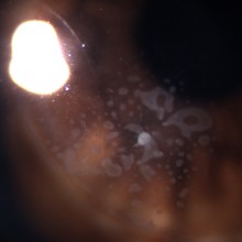
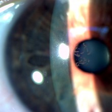
Doente de 41 anos com queixas de olho vermelho após ter estado a apanhar pinhas. Verificou-se a existência de vários pêlos de processionária no epitélio e estroma anterior da córnea. Efetuou-se desepitelização corneana com boa evolução clinica. As lesões oculares por processionária ocorrem no sul da Europa, nos meses de Fevereiro e Março.
Greasy material on eyelid surface, loss of lashes (madarosis), white lash (poliosis), eyelid margin scarring, misdirection of lashes (trichiasis), oily foamy tears, corneal marginal ulcer and inferior pannus.
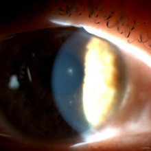
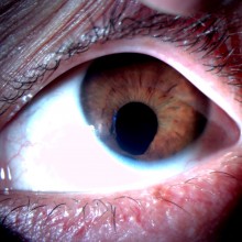
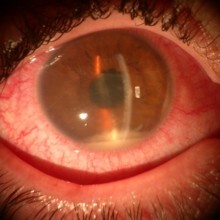
A 54-year old female patient was referred to our clinic due to long-standing progressive right eye blurred vision. No relevant personal or familial history. Right eye best-corrected visual acuity was 6/10. On biomicroscopy examination, central subepithelial spiraled opacities were characteristic of Map-dot-fingerprint Dystrophy, also known as Epithelial Basement Membrane Dystrophy or Cogan's Microcystic Epithelial Dystrophy. The disease may result in decreased vision (as in this patient) and/or recurrent corneal erosions.
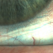
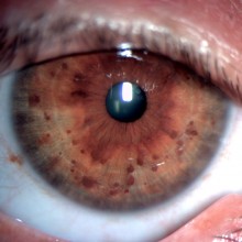
Lisch nodules on a 51-year old type 1 neurofibromatosis patient, corresponding to melanocytic hamartomas affecting the iris. The lesions are typically seen as yellowish or brown dome-shaped elevations projecting from the anterior surface of the iris.
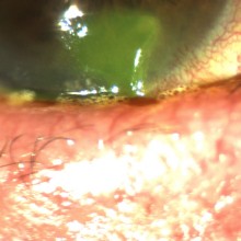
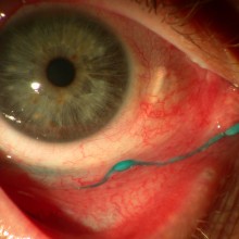
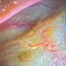
Bitot’s spots are considered pathognomic of vitamin A deficiency and correspond to triangular patches of squamous keratinous metaplasia of the conjunctival epithelium, usually near the limbus, typically at the three o’clock and/or nine o’clock positions. The foamy aspect is attributable to colonization by gas-producing bacteria (p.e. Corynebacterium xerosis).